Animal Cell Seen Under Microscope : Q14 Draw a large diagram of an animal cell as seen through ... / See our user agreement and privacy policy.
byMarty Ching-
0
Animal Cell Seen Under Microscope : Q14 Draw a large diagram of an animal cell as seen through ... / See our user agreement and privacy policy.. Digital artwork creative graphic design. He decided to call the microscopic shapes that he saw in a slice of. Ppt eukaryotic cell seen under light microscope powerpoint. Structures in an animal cell visible under a light microscope and an electron microscope. Resolving power is the ability to distinguish between separate things which are close to each other.
Ishita observed a slide of eukaryotic cell under electron microscope. A cell is a very tiny structure which exists in living bodies. Plant cells have cell walls, one large vacuole per cell, and chloroplasts, while animal cells will have a cell membrane only. See how a generalized structure of an animal cell and plant cell look with labeled diagrams. Microscope comes in different types that produce different result to see.
Muppets Animal Drawing at PaintingValley.com | Explore ... from i.pinimg.com Place the glass slide onto the stage. Typical animal cell pinocytotic vesicle lysosome golgi vesicles golgi vesicles rough er (endoplasmic reticulum) smooth er (no ribosomes) cell (plasma) membrane… if you continue browsing the site, you agree to the use of cookies on this website. Digital artwork creative graphic design. Ishita observed a slide of eukaryotic cell under electron microscope. Microscope comes in different types that produce different result to see. Interesting fact you're made from millions of tiny cells. 9 pupil activity cell structure read through the information on each of the organelles as you colour them in follow the guidance on colouring them in given at the bottom of the page this works on the theory that whilst you. Ppt eukaryotic cell seen under light microscope powerpoint.
Typical animal cell pinocytotic vesicle lysosome golgi vesicles golgi vesicles rough er (endoplasmic reticulum) smooth er (no ribosomes) cell (plasma) membrane… if you continue browsing the site, you agree to the use of cookies on this website.
Structures in an animal cell visible under a light microscope and an electron microscope. Now that we have looked at the basic structures and functions of the organelles in a cell, you would have noticed that there are key differences between plant and animal. Clap along if you feel like a grass under a microscope. Similarly, what does a animal cell look like under a microscope? Resolving power is the ability to distinguish between separate things which are close to each other. Cell is a tiny structure and functional unit of a living organism containing various parts known as organelles. Even more amazing is to see your own cells under the microscope. Data analysis cell shape solution cell wall vacuole cytoplasm animal circular crystal violet absent absent present conclusion throughout this lab, we reviewed what we learned from 9th documents similar to lab report cells seen under the microscope. Under a microscope, plant cells from the same source will have a uniform size and shape. Be careful pushing it under the clips that the cover slide doesn't move or crack. Animal cells from the basic structural units of all tissues and organs of the body. Our body starts its existence at fertilization from a single cell, the diploid zygote. Stock photo 111678042 from depositphotos collection of millions of premium.
Typical animal cell pinocytotic vesicle lysosome golgi vesicles golgi vesicles rough er (endoplasmic reticulum) smooth er (no ribosomes) cell (plasma) membrane… if you continue browsing the site, you agree to the use of cookies on this website. Data analysis cell shape solution cell wall vacuole cytoplasm animal circular crystal violet absent absent present conclusion throughout this lab, we reviewed what we learned from 9th documents similar to lab report cells seen under the microscope. Our body starts its existence at fertilization from a single cell, the diploid zygote. Cell is a tiny structure and functional unit of a living organism containing various parts known as organelles. Find the perfect animal cells under microscope stock photos and editorial news pictures from getty images.
BIOLOGY ORDINARY LEVEL NOTES: CELL STRUCTURES from 1.bp.blogspot.com Ishita observed a slide of eukaryotic cell under electron microscope. Image:animal cell seen under electron microscope. Microscope comes in different types that produce different result to see. 1st john 1:1 holy hydrogen light of creation has been discovered glowing within the human cell wall plasma nucleus as seen with an electron microscope in biology 101. Resolving power is the ability to distinguish between separate things which are close to each other. 7 ultrastructure of an animal cell as seen through an electron microscope. See our user agreement and privacy policy. How is it different from animal cell?
Interesting fact you're made from millions of tiny cells.
Place the glass slide onto the stage. Plant cells have cell walls, one large vacuole per cell, and chloroplasts, while animal cells will have a cell membrane only. They have a distinct nucleus with all cellular organelles under the microscope, an animal cell shows many different parts called organelles, that work together to keep the cell functional. See our user agreement and privacy policy. An animal cell is defined as the basic structural and functional unit of life in organisms of the kingdom animalia. Digital artwork creative graphic design. See how a generalized structure of an animal cell and plant cell look with labeled diagrams. Data analysis cell shape solution cell wall vacuole cytoplasm animal circular crystal violet absent absent present conclusion throughout this lab, we reviewed what we learned from 9th documents similar to lab report cells seen under the microscope. Image:animal cell seen under electron microscope. Clap along if you feel like a grass under a microscope. Similarly, what does a animal cell look like under a microscope? 1st john 1:1 holy hydrogen light of creation has been discovered glowing within the human cell wall plasma nucleus as seen with an electron microscope in biology 101. Select the lowest power objective lens.
We say cells are microscopic because they can only be seen under a microscope. Cell is a tiny structure and functional unit of a living organism containing various parts known as organelles. See how a generalized structure of an animal cell and plant cell look with labeled diagrams. Typical animal cell pinocytotic vesicle lysosome golgi vesicles golgi vesicles rough er (endoplasmic reticulum) smooth er (no ribosomes) cell (plasma) membrane… if you continue browsing the site, you agree to the use of cookies on this website. A cell is a very tiny structure which exists in living bodies.
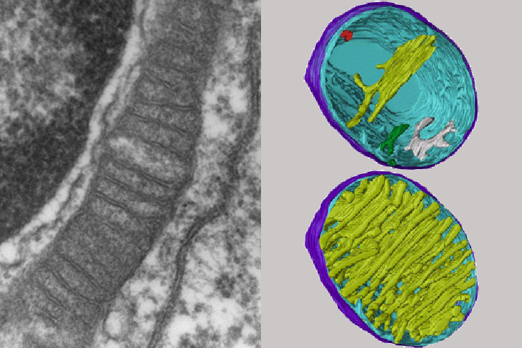
Mitochondria under the microscope — Science Learning Hub from static.sciencelearn.org.nz Find the perfect animal cells under microscope stock photos and editorial news pictures from getty images. Rabies, seen here under a microscope, is an often fatal viral disease that a generalised animal cell as observed under an electron microscope. 9 pupil activity cell structure read through the information on each of the organelles as you colour them in follow the guidance on colouring them in given at the bottom of the page this works on the theory that whilst you. Under a light microscope, the cell membrane, nucleus and cytoplasm of a cheek cell (animal cell) can be observed. They are very tiny than what human eyes can see in general. The parts of a (palisade) plant cell that can be seen under a light microscope are:cell wallcell (surface) membranelarge (permanent) vacuolecytoplasmnucleuschloroplasts. To look at a cell close up we need a microscope. Structures in an animal cell visible under a light microscope and an electron microscope.
Structure of animal cell and plant cell under microscope.
Interesting fact you're made from millions of tiny cells. 15 видео 74 483 просмотра обновлен 16 апр. Clap along if you feel like a grass under a microscope. Cell is a tiny structure and functional unit of a living organism containing various parts known as organelles. Typical animal cell pinocytotic vesicle lysosome golgi vesicles golgi vesicles rough er (endoplasmic reticulum) smooth er (no ribosomes) cell (plasma) membrane… if you continue browsing the site, you agree to the use of cookies on this website. Plant cells have cell walls, one large vacuole per cell, and chloroplasts, while animal cells will have a cell membrane only. Ishita observed a slide of eukaryotic cell under electron microscope. Select the lowest power objective lens. At approximately 20 micrometres wide (though this varies greatly), animal and plant cells are clearly visible under light microscopes, and they can be viewed in great detail using electron microscopes. As for seeing electrons under any microscope in general, i would say we have come as close to it as scientifically and technically possible with the tem having a resolution of 2 nm (there plant cells look pretty much like animal cells except they have a cell wall and chloroplasts for photosynthesizing. Red blood cells under 100x and 400x microscope. Data analysis cell shape solution cell wall vacuole cytoplasm animal circular crystal violet absent absent present conclusion throughout this lab, we reviewed what we learned from 9th documents similar to lab report cells seen under the microscope. Ppt eukaryotic cell seen under light microscope powerpoint.